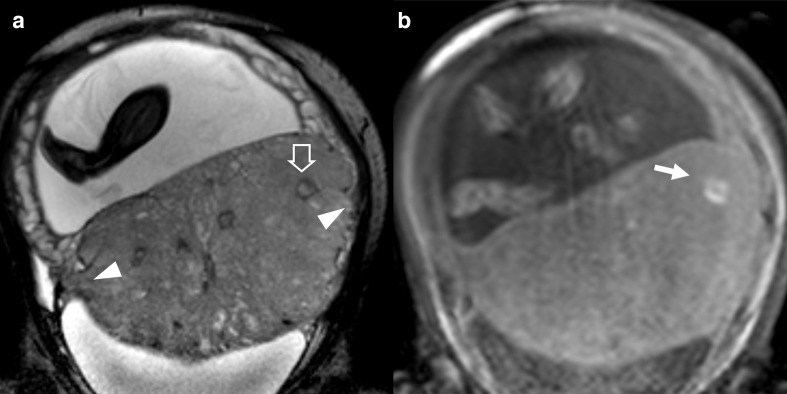
Figure 4.
patient n° 18, 32 y/o at 35th week, at third pregnancy, with two previous CS. Placenta previa percreta histologically confirmed. (A) BTFE axial and B) T1 GE coronal images. (A) penetration of the placental tissue through the myometrium which results interrupted (white arrowhead) and intraplacental nodular black band (white empty arrow) which correspond in B) to an hyperintense T1 spot (white arrow) representing a focus of haemorrhage typical of abnormal placentation. BTFE, balanced turbo field-echo.

