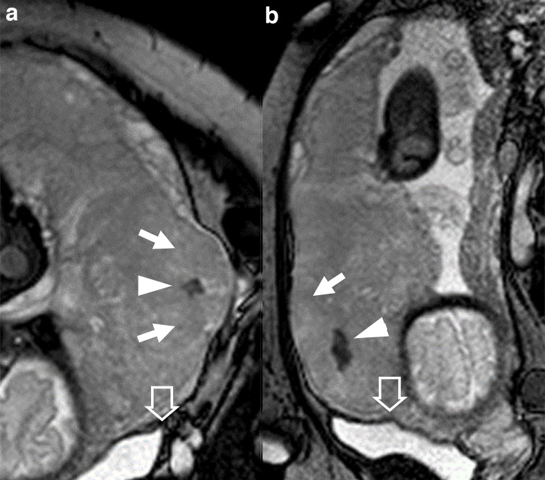
Figure 6.
patient 10, 26 y/o at 39th week, at third pregnancy, with two previous CS. Marginal placenta previa percreta histologically confirmed. BTFE images A) coronal, (B) sagittal. Big placental bulge (arrows), nodular T2 dark band (arrowheads), tenting of the bladder (empty arrows). BTFE, balanced turbo field-echo.

