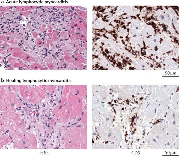
Fig. 3. Diagnosis of lymphocytic myocarditis.
Acute and healing lymphocytic myocarditis is diagnosed with histology and immunohistology of endomyocardial biopsy samples. a | Acute lymphocytic myocarditis caused by enterovirus A71 infection. Histology image showing cardiomyocyte necrosis (as revealed by the haematoxylin and eosin (H&E) staining in the left panel)) and immunohistology image showing diffuse infiltration of CD3+ T cells (as shown by anti-CD3 antibody staining (brown) in the right panel). b | Healing lymphocytic immune-mediated myocarditis. Histology image showing fibrosis but no cardiomyocyte necrosis (left panel) and immunohistology image showing the presence of infiltrated CD3+ T cells (right panel). All images ×400.

