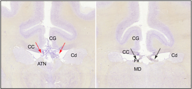
Figure 6.
Photomicrographs showing coronal sections for 1 monkey in the Experimental Group. Left, Photograph represents the selective fornix transection (red arrows indicate interaural section, 12.90 mm) above the anterior thalamus. Right, In a posterior section (interaural section, 10.90 mm), the fornix is intact, while the CC is still split. ATN, Anterior thalamus; CC, corpus callosum; Cd, caudate; CG, cingulate cortex; Fx, fornix; MD, mediodorsal thalamus.

