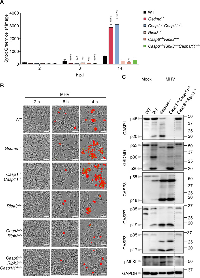
Figure 4.
Increased cell death in the absence of NLRP3/pyroptosis depends on caspase-8 and RIPK3. A, real-time analysis of cell death in BMDMs using the IncuCyte imaging system and SYTOX Green nucleic acid staining after infection with MHV. Quantification of the cell death at the indicated time points is shown. B, representative cell death images from (A) are shown. The red denotes the dead cells counted during the analysis. Scale bar, 50 µm. C, immunoblot analysis of pro- (p45) and cleaved CASP1 (p20), pro- (p53), activated (p30), and inactivated (p20) GSDMD, pro- (p55) and cleaved CASP8 (p18), pro- (p35) and cleaved CASP7 (p19), pro- (p35) and cleaved CASP3 (p17), and pMLKL in BMDMs after MHV infection for 12 h. GAPDH was used as the internal control. Data are shown as mean ± S.E. (error bars) (A). Significant differences compared with WT are denoted as *p < 0.05, **p < 0.01, ***p < 0.001, and ****p < 0.0001 (one-way ANOVA). Data are representative of at least three independent experiments. hpi, hours post-infection.

