Figure 2.
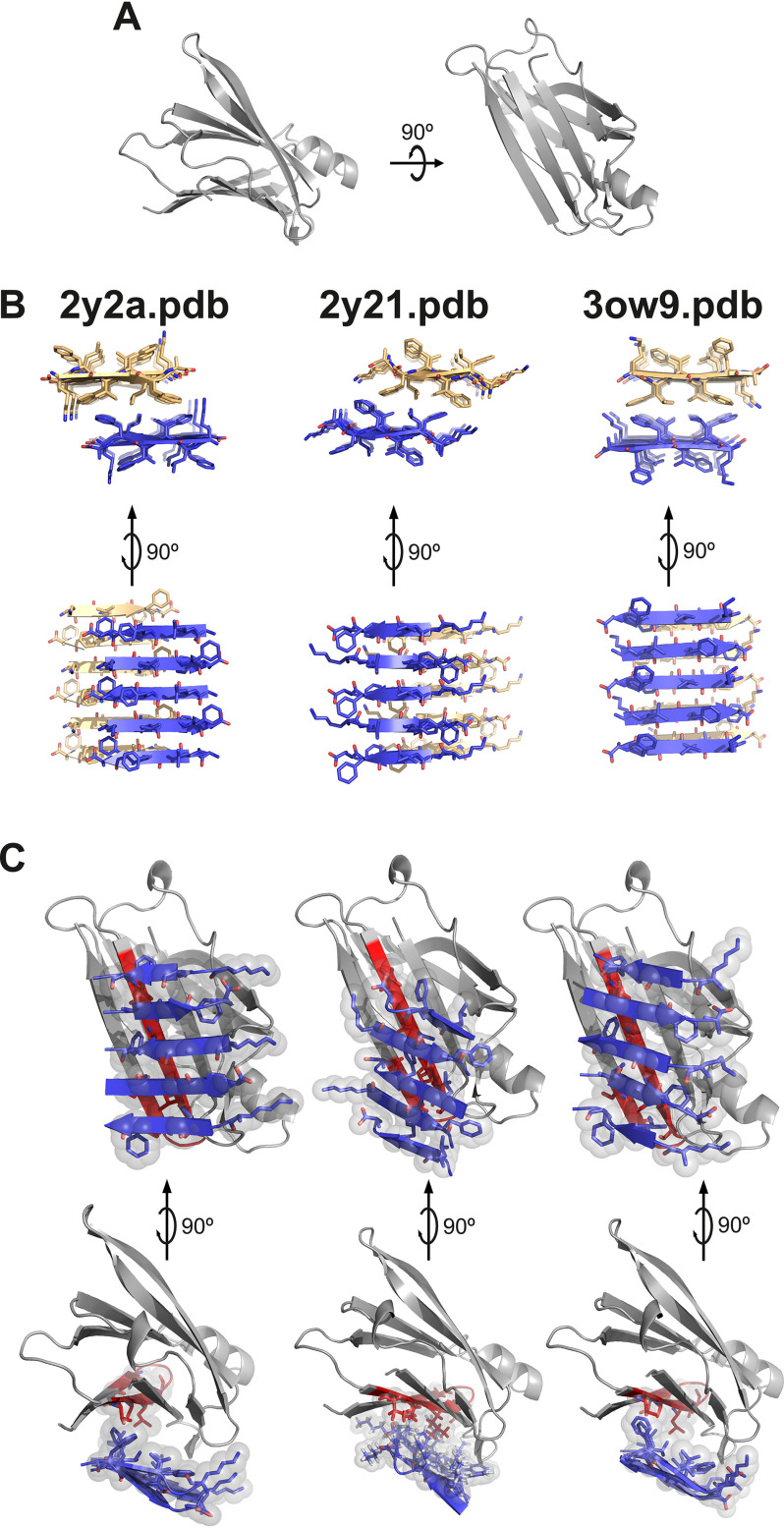
Protein–protein docking of TTR monomer with three KLVFFA polymorphs. Protein–protein docking was performed using monomeric TTR and three KLVFFA polymorphs identified previously (17): PDB codes 2y2a, 2y21, and 3ow9. A, monomeric TTR obtained from chain A of the model of PDB code 4TLT (18) is shown in gray as a secondary structure. On the left is shown a view down the hydrophobic pocket. On the right is a lateral view. B, fibrillar structures of KLVFFA from PDB codes 2y2a (left panel), 2y21 (middle panel), and 3ow9 (right panel). Only one sheet of each KLVFFA polymer was included in the docking, shown in blue. The resulting docking models, shown in C, suggest that TTR may interact with Aβ42 through residues 105–117. Monomeric TTR is shown in gray with the segment TTR(105–117) in red. Top row, lateral view of interface. Bottom row, view down the binding interface. Residues involved in the interaction between monomeric TTR and KLVFFA are shown as sticks. Spheres represent the van der Waals radii of the side chain atoms of the tightly packed binding interface.

