
Figure 5.
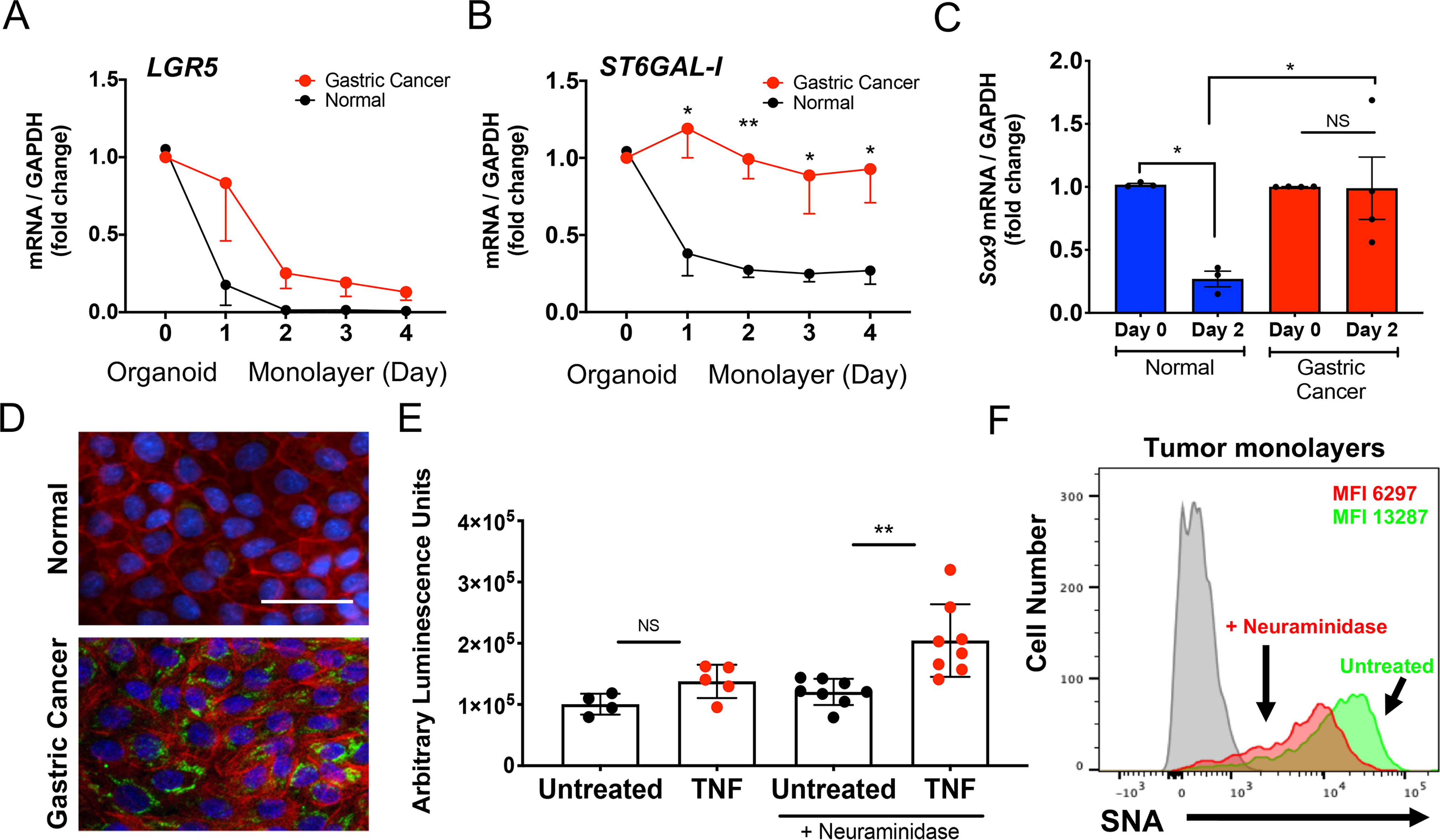
Impact of ST6Gal-I expression on gastric cancer cell apoptosis. A and B, gastric organoids and organoid-derived epithelial monolayers from gastric adenocarcinoma (red line) and normal gastric mucosa (black line) were analyzed on day 0 (organoids) and days 1–4 (epithelial monolayers) for (A) LGR5 and (B) ST6GAL1 gene expression (each n = 4). C, gastric organoids (day 0) and organoid-derived epithelial monolayers (day 2) from normal gastric tissue or gastric cancer were analyzed for SOX9 gene expression (each n = 3). D, epithelial cell monolayers generated from normal antrum-derived (top panel) and gastric adenocarcinoma–derived (bottom panel) organoids were analyzed on day 2 by immunofluorescence by staining with an ST6Gal-I antibody (FITC), phalloidin (Alexa Fluor 594) and DAPI (representative donor, 20×; n = 4; scale bar, 50 μm). E, gastric cancer organoid–derived epithelial monolayers were pretreated with or without A. ureafaciens neuraminidase and assayed for caspase 3/7 activity (n = 4–8 in three separate gastric cancer-derived monolayers). F, gastric organoid–derived epithelial monolayers were analyzed for SNA expression using flow cytometry with (red) or without (green) A. ureafaciens neuraminidase pretreatment. A–C, E, significance: *, p < 0.05; **, p < 0.01; ***, p < 0.001; and ****, p < 0.0001.