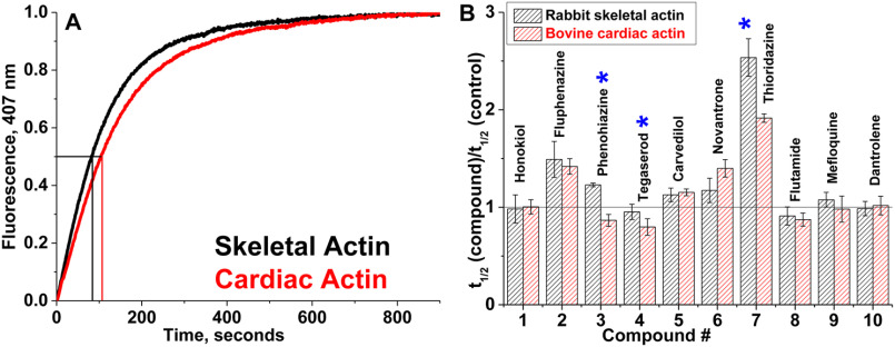
Figure 3.
Actin polymerization. A, time courses of polymerization of skeletal and cardiac actin in the absence of compounds. The increase in fluorescence of skeletal and cardiac actin, containing 5% pyrene actin, was monitored at 407 nm. Polymerization half-times (vertical lines) denote the times required for actin to reach half-maximal fluorescence (horizontal line). For each actin species, at least three independent preparations were used. B, relative change in apparent half-times (t½) of polymerization of actin isoforms in the presence of 50 μm compound. Errors are reported as standard deviation, n = 3. Significant difference (*) in drug effect on skeletal versus cardiac actin was established with Student's t test, p < 0.05.

