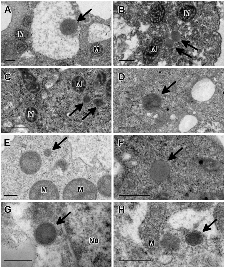
Fig. 1.
Identification of peroxisomes in three genera of pathogenic free-living amoebae by transmission electron microscopy. Spherical structures with a dark granular content and limited by a single membrane were observed in Acanthamoeba griffini (A), Acanthamoeba polyphaga (B), Acanthamoeba royreba (C), Acanthamoeba castellanii (D), Balamuthia mandrillaris (E), and Naegleria fowleri (F). These structures ranged in size from 0.2 to 0.9 µm and they were morphologically similar to peroxisomes from mouse liver samples, which are shown for comparison (G, H). M, mitochondrion; Nu, nucleus. Bar = 0.5 µm.

