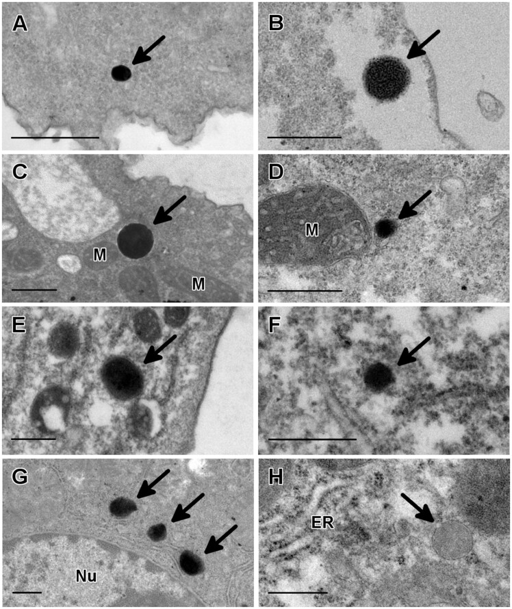
Fig. 2.
Peroxisome identification in three genera of pathogenic free-living amoebae by cytochemical staining. Thin sections of trophozoites were treated with diaminobenzidine and hydrogen peroxide to detect catalase activity. Positive reaction products were clearly seen in Acanthamoeba griffini (A), Acanthamoeba polyphaga (B), Acanthamoeba royreba (C), Acanthamoeba castellanii (D), Balamuthia mandrillaris (E) and Naegleria fowleri (F). In all of these amoebae, the labeling was deposited in round structures with a uniform electrondense content. Mouse liver samples were used as a positive control for the cytochemical staining (G). As a negative control for the cytochemical reaction, a mouse liver sample was incubated without H2O2 (H). ER, endoplasmic reticulum; M, mitochondrion; Nu, nucleus. Bar = 0.5 µm.

