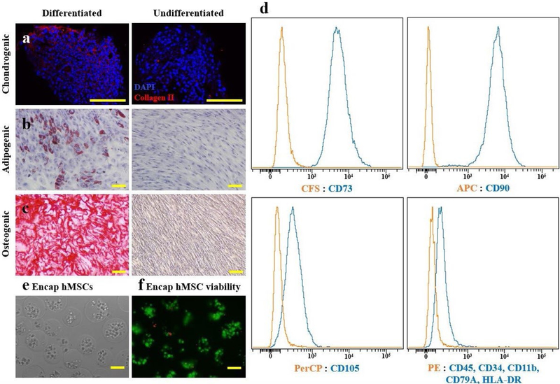
Fig. 2. Encapsulated hMSC characterization.
Multipotency of hMSCs confirmed prior to encapsulation, as cultured hMSCs were differentiated into (a) chondrogenic, (b) adipogenic and (c) osteogenic phenotypes as demonstrated by collagen type II, oil red O and alizarin red staining. (d) FACS analysis demonstrated that hMSCs expressed typical MSC surface markers: CD73, CD90 and CD105; but not hematopoietic markers: CD45, CD34, CD11b, CD79A and HLA-DR. (e) Light microscopic appearance of encapsulated hMSCs immediately following sodium alginate encapsulation showed capsule diameters of 170 ± 27 μm. (f) Fluorescent viability assay, staining live cells green and dead cells red, showed 96 ± 2.4 % of cells were viable immediately following encapsulation in sodium alginate. Scale bars = 100 μm.

