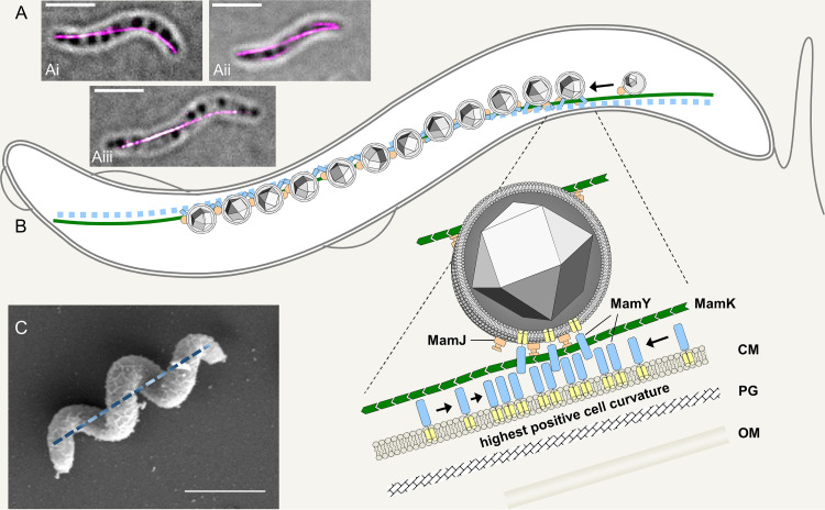
FIG 3.
Localization and function of magnetoskeleton constituents. (A) Localization of mCherry-MamK (i and ii) and mCherry-MamY (iii) in live M. gryphiswaldense cells imaged by 3D structured illumination microscopy (3D-SIM; maximum-intensity projection overlaid with a bright-field image; scale bar, 2 μm). (B) 2D model of the current view of the “magnetoskeleton” in M. gryphiswaldense, as suggested by Toro-Nahuelpan et al. (99). The actin-like MamK (green) polymerizes into cell-spanning dynamic cytoplasmic filaments that nucleate at the cell poles and “treadmill” toward midcell. Maturating magnetosomes become attached to this filament via MamJ (orange). MamY is a protein of the cytoplasmic and magnetosome membranes (membrane helices, yellow; cytoplasmic domain, blue) with high potential to self-interact. MamY assemblies are suggested to become curvature sensitive and localize to sites of highest positive inner membrane curvature coinciding with the geodetic cell axis, where they recruit magnetosome chains. CM, cytoplasmic membrane; PG, peptidoglycan; OM, outer membrane. The model is not drawn to scale. (C) SEM image of an M. gryphiswaldense cell illustrating a corkscrew-like cell morphology. The geodetic cell axis is indicated as a dashed line. Scale bar, 2 μm.

