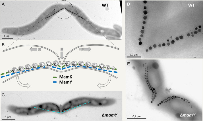
FIG 4.
Cytokinesis in the presence of a magnetosome chain, as suggested by Katzmann et al. (94). (A) TEM image of a dividing M. gryphiswaldense wild-type cell. The center of the magnetosome chain is precisely localized at the division plane, ensuring that each daughter cell inherits exactly half of the magnetosomes (89, 93). Note the “buckling” cell center reflecting a distortion of the otherwise preserved helical cell morphology. (B) 2D scheme of the center from a dividing wild-type cell. Shaded arrows indicate direction of main cell wall growth at the division plane. Open arrows indicate the direction of cell bending. (In three dimensions, some twisting may occur as well.) Within the division plane, the septum grows by asymmetric indentation starting unilaterally from the site of negative inner cell curvature, resulting in a fracture-like appearance of the magnetosome chain. (C) In the mamY mutant, the magnetosome chain seemingly becomes disrupted by the wedge-like growing division septum. However, the buckling cell shape upon division is still apparent. The dashed line indicates the magnetosome chain position in wild-type cells. (D and E) Magnification of magnetosome chains immediately after splitting in the wild type (D) and the mamY mutant (E).

