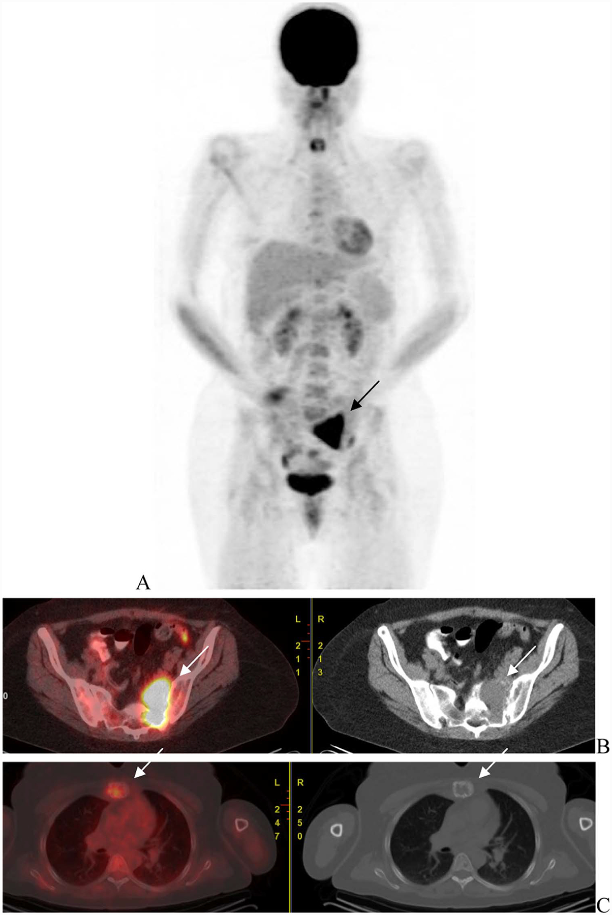
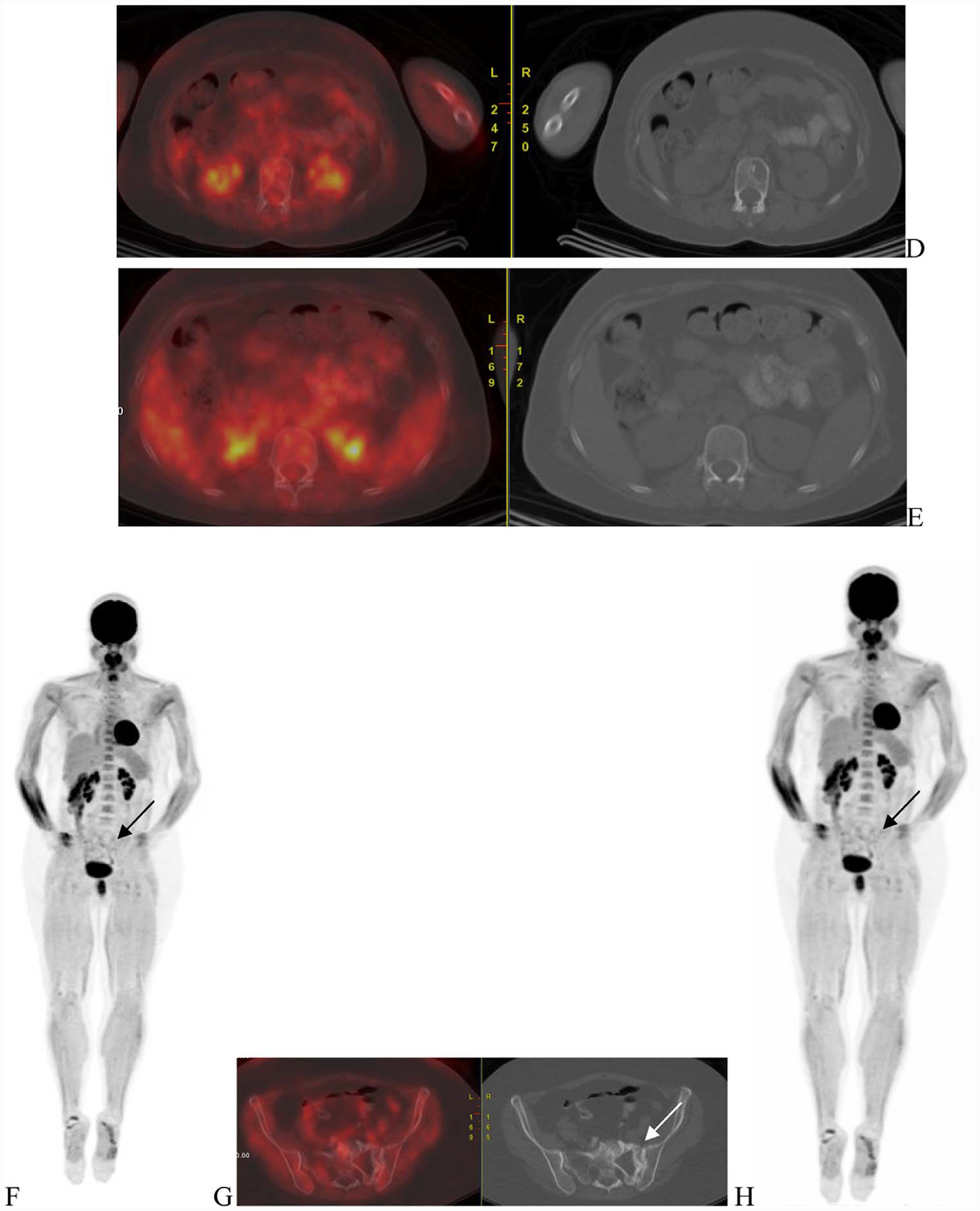
Fig. 3.


A 48-year-old woman with ISS-1 disease. FGD PET scan was done at baseline that showed uptake in the sacrum (A—arrow) corresponding to a large lytic lesion in noted on the CT with soft tissue component (B—arrow). Mild uptake was nted in the sternal lesion (C—arrows). Other lytic lesions were noted on the CT (D and E—arrows) but did not have corresponding increased uptake.
