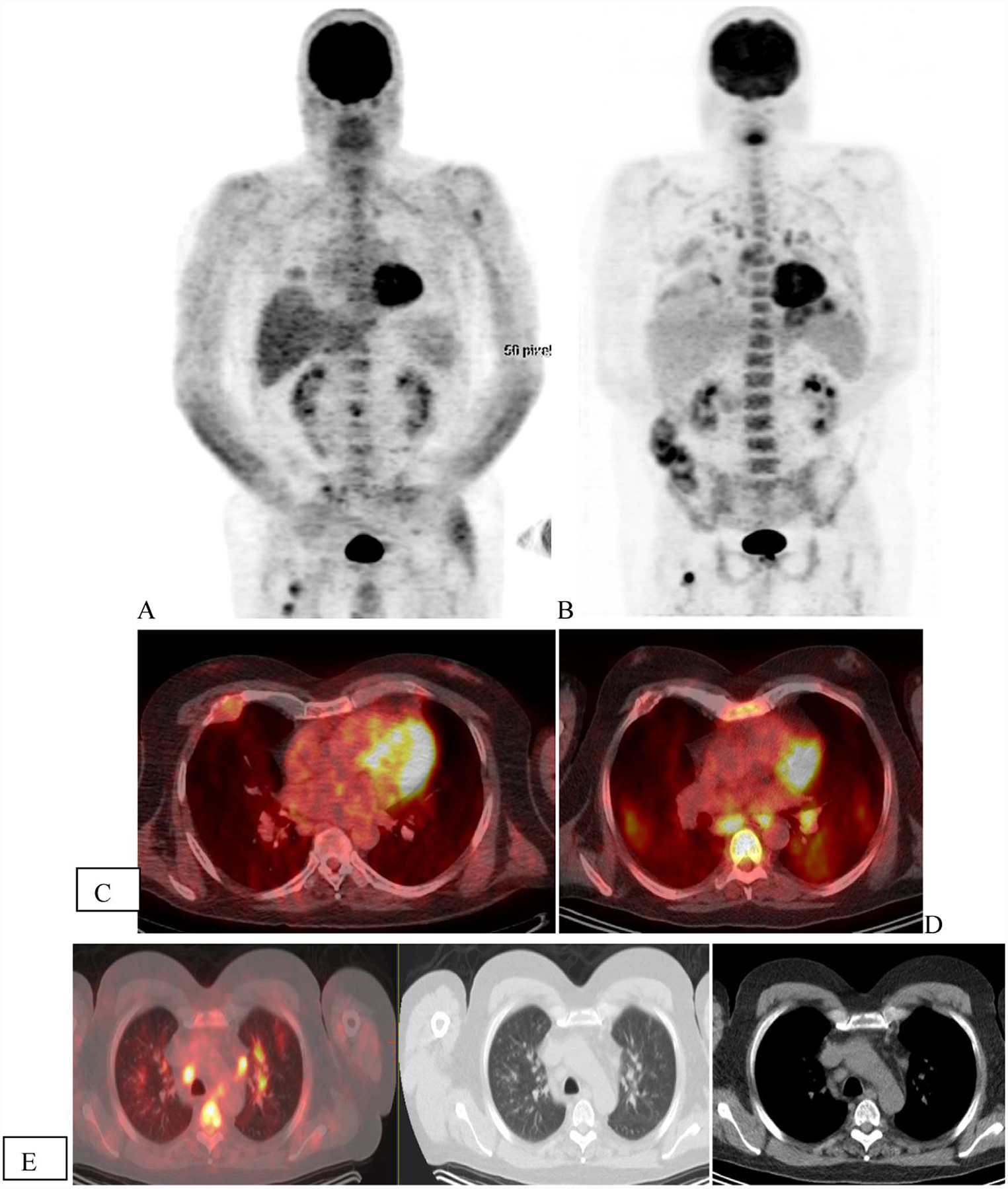
Fig. 5.

Patient with ISS2 IgA kappa multiple myeloma with high-risk disease. Initial scan (A) shows uptake in the lytic lesion in rib, vertebra and femur (arrows) that show mixed changes in FDG uptake with less uptke in the rib abd vertebra but higher uptake in the femur. Increased FDG uptake was seen in bilateral lung with infiltrative changes and mediastinal and hilar nodes (arrows) that was attributed to infectious, inflammatory change.
