
Fig. 6.
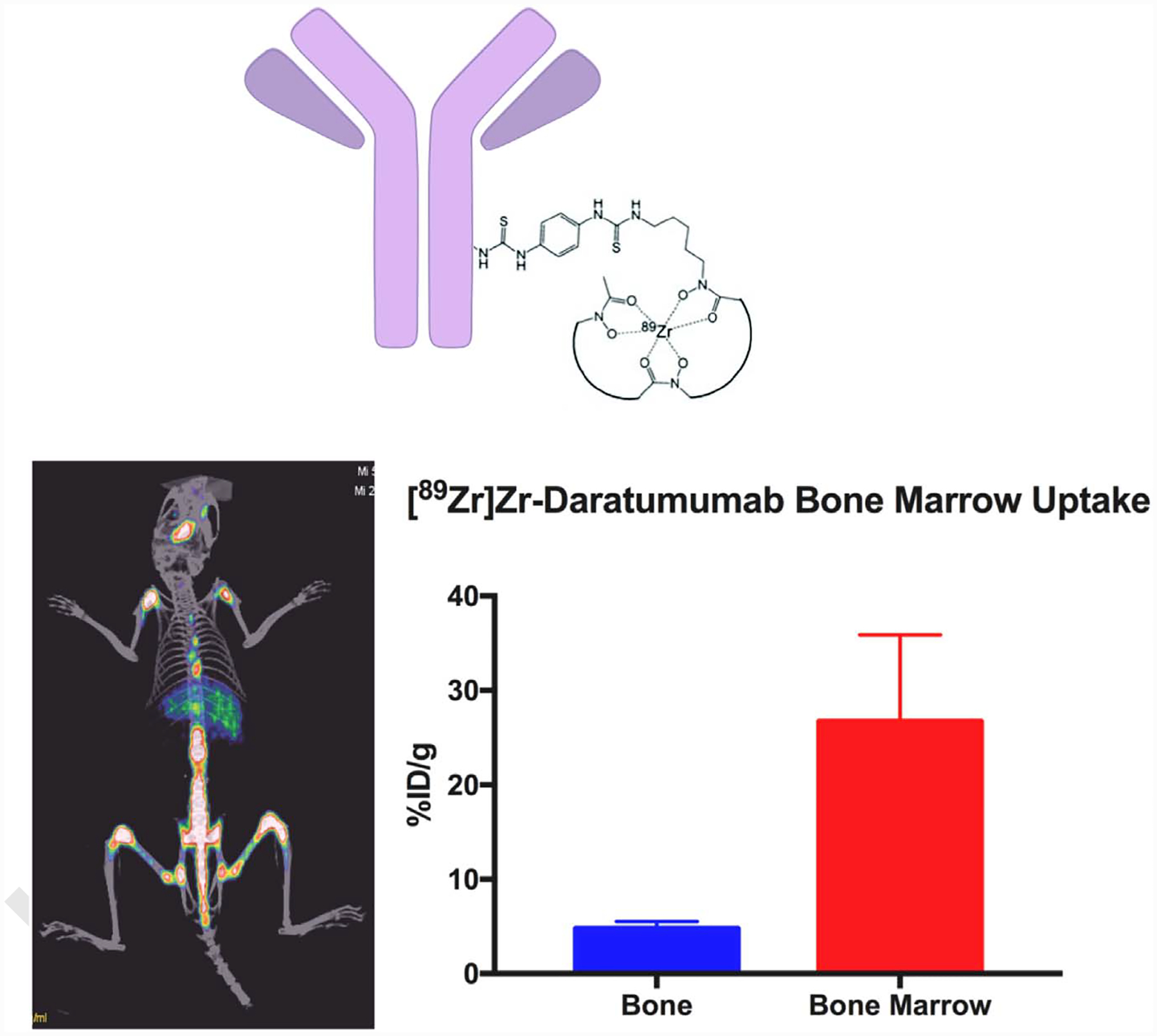
(A) Structure [89Zr]Zr-DFO-daratumumab. (B) Imaging with [89Zr]Zr-daratumumab in a disseminated mouse model (OPM-2 cells in NSG mice) of multiple myeloma (Sobol et al., unpublished data). There is prominent uptake in bones as seen along the spine and limbs. The uptake localized to the marrow and bone disease induced in the mice. This data corroborate the work done by the Shokeen group at Washington University (Ghai et al., JNM 2017) in a different MM model, showing that [89Zr]Zr-daratumumab has the ability to noninvasively identify MM in mouse models. This technology is currently being translated to the clinic at MSK. (Courtesy: Nicholas Sobol, PhD and Jason Lewis, PhD, Radiochemistry and Molecular Imaging Probes Core, MSK).