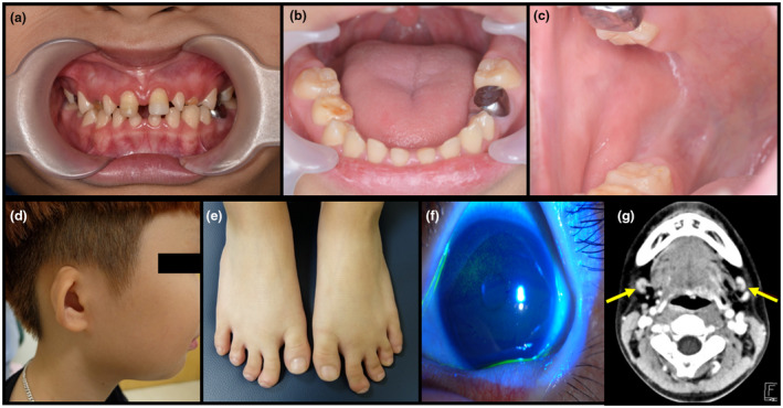
Figure 1.

Clinical photographs and radiologic study of the proband. Proband displays severe xerostomia, multiple caries, enamel hypoplasia, microdontia, missing teeth, and a bald tongue (a and b). The orifice of the Stensen's duct is unidentifiable (c). He has low‐set, cup‐shaped ears (d). He has clinodactyly in the second and third toes of both feet (e). Slit lamp examination reveals punctate epithelial erosions on the cornea due to xerophthalmia (f). Computed tomography of the neck shows no visible parotid glands, with small enhancing nodular lesions in both sides of the neck, level IB (g)
