
Figure 2.
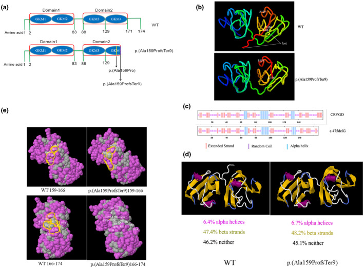
Bioinformatics analysis of the mutant CRYGD p.(Ala159ProfsTer9). (a) Diagrammatic scheme of the functional domain of CRYGD and p.(Ala159ProfsTer9) protein. (b) The tertiary structure of CRYGD and p.(Ala159ProfsTer9) protein. (c) The predicted secondary structure showing the reduced extended strand and random coil in the mutant. (d) The predicted secondary structure showing the changed alpha‐helices and beta‐strands in the mutant. (e) The predicted hydrophobic surface and amino changes in the mutant