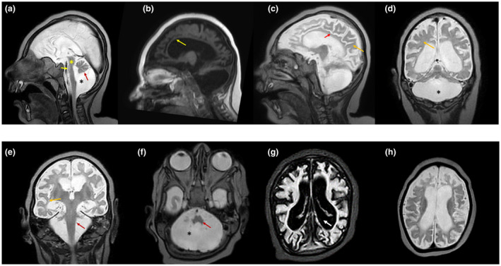
Figure 2.

Magnetic resonance imaging characteristics of the proband (V.2). (a) Sagittal cut through the midline (T2‐W) shows the loss of transverse fibers of the pons (yellow asterisk), mega cisterna magna (black asterisk), hypoplasia of brainstem, cingulate gyrus, pons, vermis and cerebellar hemispheres (black arrow). The findings described would be highly compatible with pontocerebellar hypoplasia (black asterisk). (b) The corpus callosum is almost absent. (c) Hypoplasia of the cingulate gyrus (red arrow) and white matter (yellow arrow) are evident. (d and e) Lateral sagittal section (T2‐W) shows hypoplasia of the cerebellar hemispheres (asterisk). The white matter loss (delayed myelination) is also evident (yellow arrow). The red arrow shows the almost complete hypoplasia of the cerebellum. (f) Hypoplasia of cerebellum (asterisk) and pons (red arrow) is evident. (g) The coronal section (T2‐W) shows extremely small cerebellar hemispheres and extended vermal hypoplasia. Immaturity of the cerebral cortex and ventriculomegaly (white thick arrow) are also clearly evident. (h) Coronal sections show hypoplasia of the cerebellar hemispheres. An enlarged ventricle is visible and also there is an increased distance between the cortical surface and the skull evident, which is probably due to diminished brain growth in utero (IUGR)
