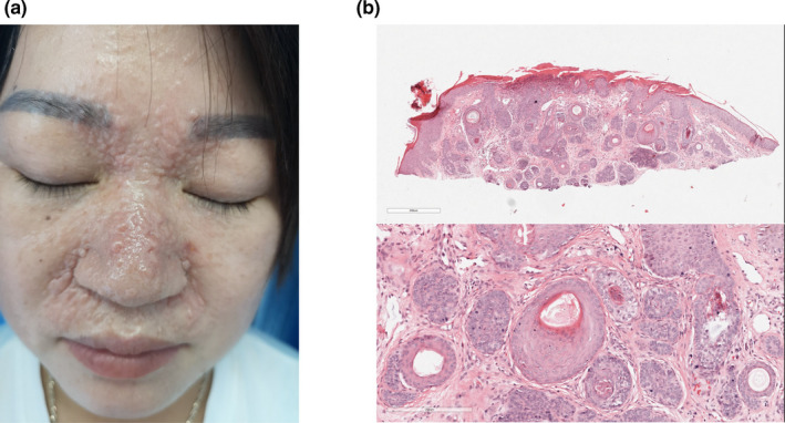
Figure 2.

Clinical and histopathological features of the proband. (a) The picture showed the proband with multiple discrete and confluent skin‐colored papules and nodules located on the face, especially in nasolabial folds and inner aspects of eyebrows. (b) The figure showed the histopathological features of the proband. The neoplasm was composed of several fibroepithelial units, in which basaloid cells formed in a fibrous stroma with follicular germs and papillae
