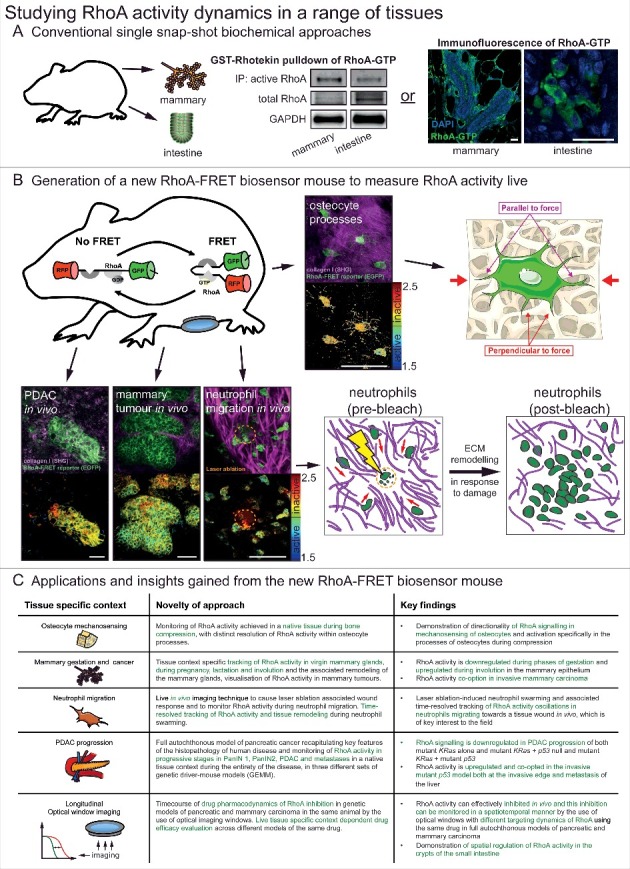
Figure 1.

Studying RhoA activity dynamics in a range of tissues. (A) Conventional single snap-shot based biochemical approaches to analysing RhoA activity in two examples tissues of the mammary gland and intestine. These included bead-based pulldown of RhoA-GTP in tissue lysate and a recently developed immunofluorescence of fixed tissue samples using a RhoA-GTP specific antibody [15,19]. scale bars, 25 μm (B) With the generation of the new RhoA-FRET biosensor mouse RhoA activity could be monitored live in osteocytes of the calvaria, in vivo in pancreatic ductal adenocarcinomas, mammary tumours and during neutrophil migration (RhoA-FRET biosensor, green; collagen-derived second harmonic generation (SHG) signal, magenta) with corresponding fluorescence lifetime imaging microscopy (FLIM) images of RhoA activity (high RhoA activity: blue to green; low RhoA activity: yellow to red). scale bars, 50 μm (C) A summary of the new insights gained by the use of the new RhoA-FRET biosensor mouse in a variety of tissues and applications. Adapted from Nobis et al. 2017, Cell Reports and adapted from Servier Medial Art, licensed under the Creative Commons Attribution 3.0 Unported license (https://creativecommons.org/licenses/by/3.0/).
