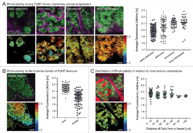
Figure 2.

Spatially defined RhoA acitivty during the progression of PyMT-driven mammary carcinomas. (A) RhoA-FRET mice crossed to MMTV-polyoma-middle-T antigen (PyMT) mice allow for the tracking of RhoA activity during the progression of invasive mammary carcinoma (n = 1 mouse, 280 cells). (B) RhoA activity is increased at the invasive borders of primary PyMT tumours (white dashed line) compared to tumour core regions (n = 1 mouse, 180 cells). (C) Intravenous injeciton of a contrast dye (Qtracker655) allows for monitoring of RhoA activity in cancer cells in relation to their proximity to local vasculature (n = 2 mice, 130 cells). Dots, single cells; line, mean; error bars, SD; scale bars, 50 µm.
