Figure 4.

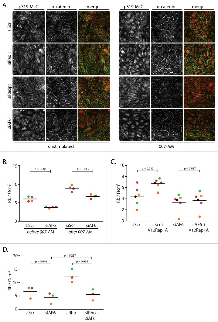
AF6 mediates Rap1-induced junctional tension and concomitant barrier function. (A) Immunofluorescence of HUVEC monolayers transfected with control siRNA (siScr), siRNA targeting Radil (siRadil), Rasip1 (siRasip1) or AF6 (siAF6), either not stimulated or stimulated with 1 µM 007-AM 15 minutes prior to fixation. The cells were stained for pS19-MLC and α-catenin. The merged image depicts pS19-MLC in red and α-catenin in green. Knockdown efficiencies are shown in supplemental figure 1E. (B) Endothelial barrier (Rb) of control HUVEC monolayers (siScr) and HUVEC monolayers depleted of AF6 (siAF6), either before or 45 minutes after stimulation with 1 µM 007-AM. Different colors represent independent experiments (n = 4). Averages are indicated by the black lines. Knockdown efficiencies are shown in supplemental figure 1F. (C) Endothelial barrier (Rb) of control HUVEC monolayers (siC) and HUVEC monolayers depleted AF6 (siAF6), transduced with control lentivirus or G12VRap1A containing lentivirus. Different colors represent independent experiments (n = 6). Averages are indicated by the black lines. Knockdown efficiencies are shown in supplemental figure 1G. (D) Endothelial barrier (Rb) of control HUVEC monolayers (siScr) and HUVEC monolayers depleted of AF6 (siAF6) and/or RhoA, RhoB and RhoC (siRho). Different colors represent independent experiments (n = 3). Averages are indicated by the black lines. Knockdown efficiencies are shown in supplemental figure 1H.
