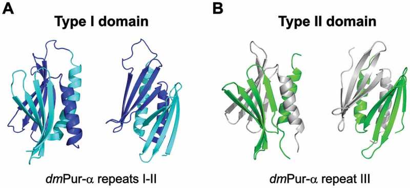
Figure 3.

Structure of Drosophila melanogaster Pur-α protein exhibiting type I (A) (PUR repeat I-II; PDB ID: 5fgp) [15] and type II (B) (PUR repeat III; PDB ID: 5fgo) [15] PC4-like fold. Each structure is shown in two different orientations. The structure of type I is represented in cyan (repeat I) and navy blue (repeat II), whereas in the structure of type II PC4-like fold individual chains are shown in green and grey. The figure has been prepared in PyMol v 1.3 (pymol.org).
