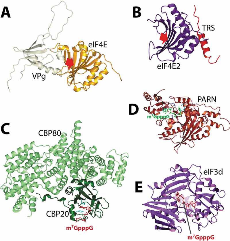
Figure 3.

Structural insights into cap substitutions and other cap-binding proteins. Panel A. NMR based model of eIF4E/VPg complex highlighting the competition with the m7G cap in the cap-binding pocket (red arrow). Panel B. eIF4E2 bound to TRS, using a similar surface used by eIF4E to bind eIF4G or 4E-BP1. The crystal structure was not solved in the presence of a cap analogue, but the cap-binding pocket is shown with the red arrow. Panels C and D. Structures of factors identified demonstrating the similar means by which they interact with the m7G cap. This same pocket is use to bind TMG cap for CBC. Panel E. eIF3d cap-binding domain modelled with the cap analogue placed according to its position in DXO. PDB codes are as follow: 5XLN (B); 1H2U (C); 3D45 (D); 5K4B (E).
