Figure 2.

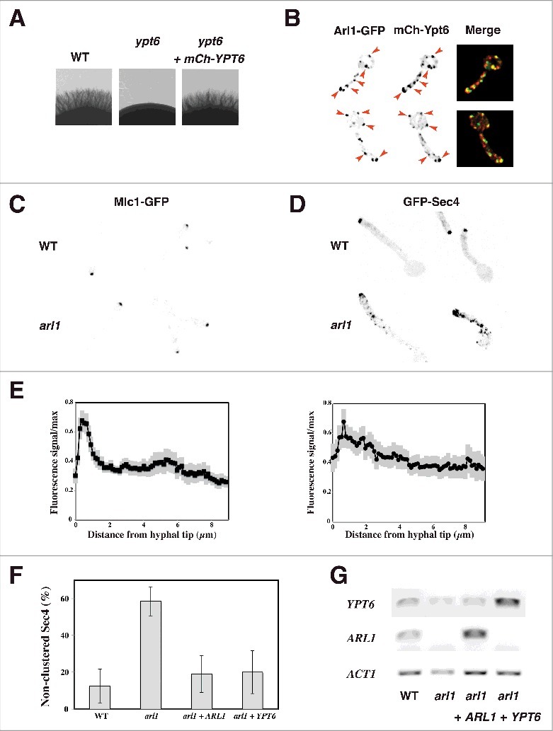
Overexpression of YPT6 rescues the altered Sec4 distribution in the arl1 deletion mutant. (A-B) Arl1 and Ypt6 partially co-localize in hyphae. The mCh-Ypt6 fusion is functional (A) Cells from the indicated strains were incubated on Spider media and images were taken after 5 days. Similar results were observed in 2 independent experiments. Maximum projections of 21 deconvolved z-sections of representative cells expressing Arl1-GFP together with mCh-Ypt6 after 90 min FCS-induced hyphal growth; GFP and mCherry signals were acquired simultaneously. (B) (C-E) Sec4, but not Mlc1, distribution is altered in arl1/arl1 cells. WT and arl1/arl1 cells, expressing Mlc1-GFP (C) or GFP-Sec4 (D), were incubated with FCS for 45 and 90 min, respectively. Representative images are shown. Cell fluorescence concentration profiles were analyzed with the HyphalPolarity program,47 to quantify the GFP-Sec4 signal concentration along the major axis of hyphal cells, from sum projection images generated with Image J (E). (F) Overexpression of YPT6 restores Sec4 clustering in arl1/arl1 cells. The graph shows averages of 3 independent experiments (n = 10–30 cells each) for each indicated strain, with p values: arl1 vs WT: 0.0004, arl1 + ARL1 vs arl1: 0.0028 and arl1 + YPT6 vs arl1: 0.0042; no statistically significant difference was observed between the values for WT, arl1 + ARL1 and arl1 + YPT6. (G) YPT6 and ARL1 transcripts in strains expressing GFP-Sec4. Transcripts were determined as in Fig. 1F.
