
Figure 4.
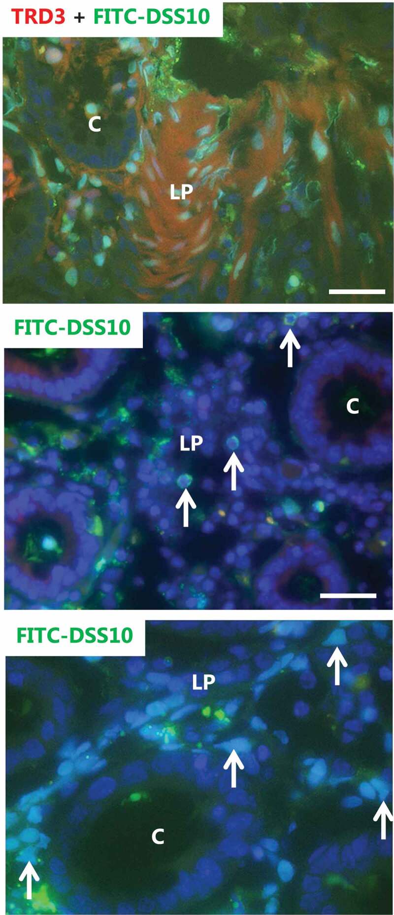
Nuclear targeting of FITC-DSS10, but not of unsubstituted FITC-dextran. Explants from pig colon were cultured for 1 h in the presence of FITC-DSS10 and TRD3. The top image shows that whereas TRD3 mainly stained the lamina propria diffusely, the FITC-DSS10 distinctly targeted nuclei. The nuclei were most often evenly labeled, but occasionally the labeling was confined to the nuclear rim (indicated by arrows in the middle image). The bottom image of increased magnification shows that FITC-DSS10-positive nuclei were sometimes of abnormal shape (indicated by arrows). Bars: 50 µm.