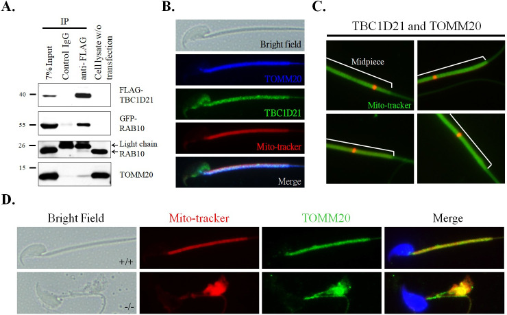
Fig 6. TBC1D21 interacts with one of the mitochondria membrane proteins, TOMM20.
(A) co-IP of FLAG-TBC1D21 with RAB10 and TOMM20. Lysates from the cells that were transfected with the pFLAG-TBC1D21 and GFP-RAB10 vector and then immunoprecipitated with a nonspecific control IgG (Lane 2), or an anti-FLAG antibody (Lane 3), followed by immunoblotting with the FLAG antibody, GFP antibody, RAB10 antibody, or TOMM20 antibody. An input protein (7%) was used as a control during the immunoblotting of the transfected cell lysates (Lane 1). (B) Immunofluorescence detection of TOMM20 was found to be co-localized with TBC1D21 in wild-type mature spermatozoa. Figures from top to bottom indicate the bright field, TOMM20 signals (blue), TBC1D21 signals (green), Mito-tracker (red), and merged figure in wild-typmature spermatozoa. (C) Evaluation of the interaction between TBC1D21 and TOMM20 in vivo in four wild-type mature spermatozoa using the Duolink PLA assay. The white lines represent the midpiece region in mature spermatozoa. The arrow indicates the localization sites of the TBC1D21/TOMM20 complexes (red dots). (D) IF assay results showing disturbed localizations of TOMM20 signals in Tbc1d21-defective sperm, compared with wild-type sperm. Figure from left to right: the bright field, Mito-tracker (red), TOMM20 signals (green), and figure merged with Mito-tracker, TOMM20, and DAPI (blue). Figures shown as wild-type sperm (upper panels) and Tbc1d21 knockout sperm (lower panels). Magnification = 1000X.

