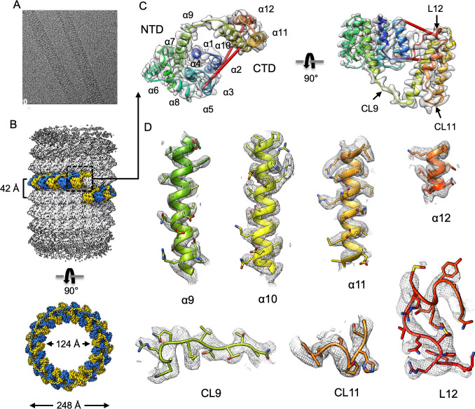
Fig 5. Cryo-EM helical reconstruction of M1-V97K filament.
(A) Representative micrograph. (B) Helical reconstruction of M1-V97K oligomers reveals a hollow tube with inner and outer diameters of 124 Å and 248 Å. One helical turn comprises approximately 21 asymmetric units, highlighted in alternating blue and yellow colors. (C) Ribbon representation of one M1-V97K asymmetric unit, which includes the NTD helices α1–9, the newly resolved CTD helices α10–12, and connecting loops CL9–11 and terminal loop L12. Identified red crosslinks are shown as red rods. (D) Cryo-EM maps and models of M1-V97K helices α9–12 and connecting loops CL9–11 and terminal loop L12. Most side chains are clearly resolved in the cryo-EM map at 3.4 Å resolution. See also S4 and S5 Figs and S3 Table. cryo-EM, cryo-electron microscopy; CTD, C-terminal domain; M1, matrix protein 1; NTD, N-terminal domain.

