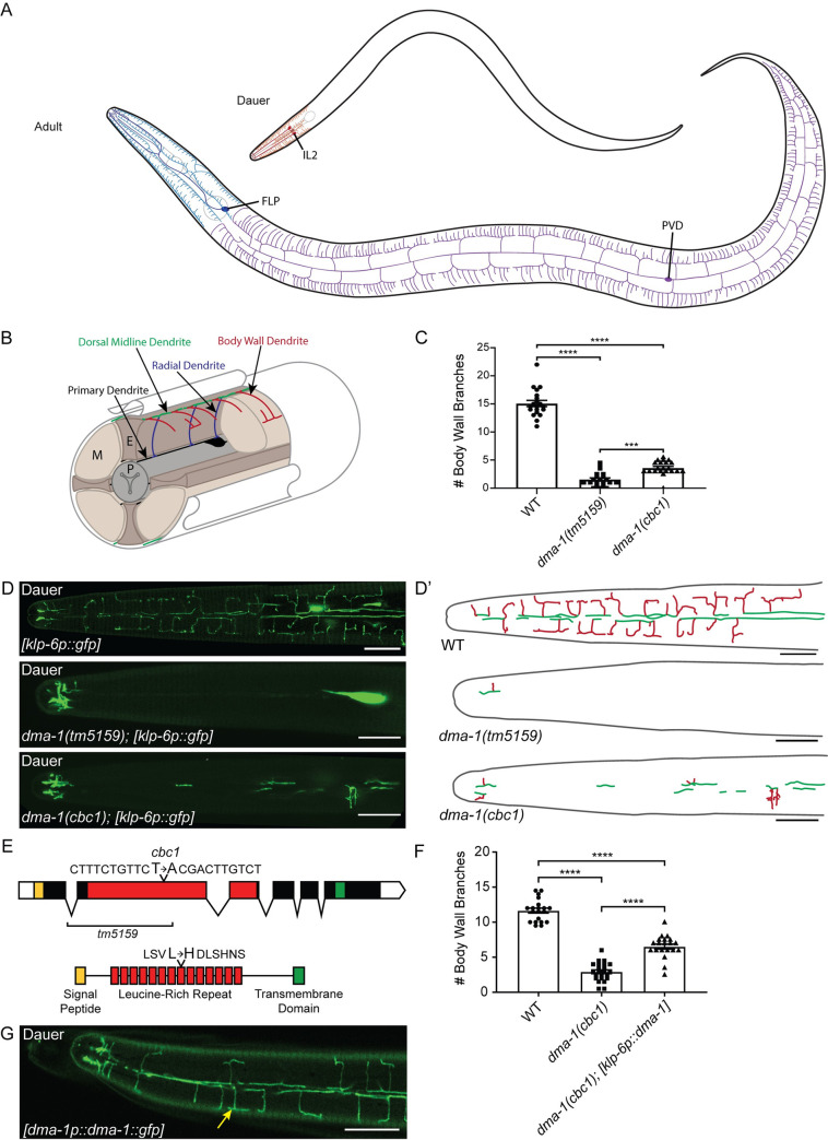
Fig 1. DMA-1 is required in the IL2s for arbor formation.
(A) There are three classes of highly arborized neurons in C. elegans: IL2, FLP, and PVD. The IL2s (red) arborize strictly during the dauer stage where their arbors are restricted to the head of the nematode. The FLPs (blue) and PVDs (purple) form arbors during late L4-adult and together cover the head and body, respectively. (B) A schematic cross-section of the IL2 branching pattern, shown in ¾ view. The IL2 cell body and primary dendrite (established during embryonic development) are black. Subsequent dendritic growth occurs during dauer formation leading to branches from the primary dendrites towards the ventral and dorsal midlines in a radial arrangement (blue), branches along the midlines (green), and body wall branches extending from the midlines (red). P = pharynx, E = epidermis, M = muscle. (C) Quantification of the number of IL2 branches along the body wall in wild-type (n = 20), dma-1(tm5159) (n = 21), and dma-1(cbc1) (n = 21). (D) Z-projection confocal micrographs of IL2 body wall branches in dauers. dma-1(cbc1) mutants have severe defects in IL2 arborization. The deletion allele, dma-1(tm5159), has a greater reduction of branching than the dma-1(cbc1) allele. The IL2 neurons are labeled with klp-6p::gfp. (D’) Schematic drawings of IL2 arbors traced from the accompanying micrographs illustrating the dorsal midline (green) and body wall branches (red). (E) Gene schematic of dma-1 (top). dma-1(cbc1) is a point mutation that results in a T to A transversion within the leucine-rich repeat (red) region. The previously described deletion allele dma-1(tm5159) removes much of the LRR domain (9). Protein domain schematic (bottom) highlights the leucine to histidine mutation in dma-1(cbc1) within the eighth leucine-rich repeat. (F) Quantification of the number of IL2 branches along the body wall during dauer in wild-type (n = 19), dma-1(cbc1) (n = 18), and dma-1; klp-6p::dma-1 (n = 20). Expression of dma-1 in the IL2 neurons using the klp-6 promoter partially rescues the dma-1 mutant branching phenotype. (G) Z-projection confocal micrograph of a dauer expressing dma-1::gfp under the dma-1 promoter. dma-1p::dma-1::gfp localizes to the IL2 dendrites (yellow arrow) during dauer. This image substack includes the dendrites located at the body wall. For IL2 body wall branch quantification (C and F), an ANOVA followed by Tukey’s test for multiple comparisons was used to determine significance, ***p ≤0.001 and ****p ≤0.0001. Error bars, standard error of the mean. Scale bars, 10 μm.

