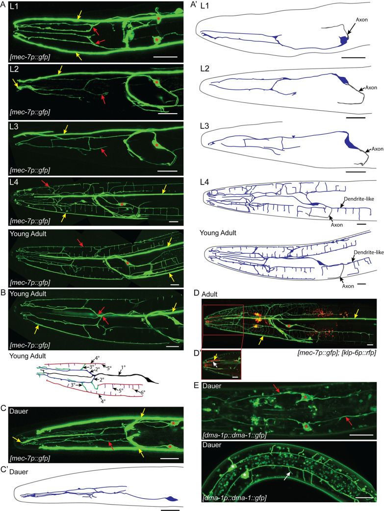
Fig 3. FLP branching architecture.
(A) Z-projection confocal micrographs of FLP neurons throughout larval development. Dorsal-ventral view of the FLP dendrite in L1. Each FLP primary dendrite ranges from the cell body to anterior of the metacorpus where it branches to send processes towards the nose. Lateral views of the L2, L3, and L4 FLP dendrite with the focal plane centered around the left FLP arbor. Little additional branching occurs during L2 and L3. The FLPs arborize rapidly during L4. Lateral view of the adult FLP arbors showing extensive dendritic arborization covering the head of the nematode. The primary dendrite travels along the lateral sensory neuron fascicle until anterior of the metacorpus. There it branches and sends secondary processes to the subdorsal and subventral sensory neuron fascicles. A thin primary dendrite continues within the lateral sensory fascicle towards the nose. The secondary dendrites in the subventral and subdorsal fascicles branch again to send processes to the dorsal or ventral midlines and sublateral lines along the body wall where they branch to form the body wall candles of the arbor. Red arrows indicate part of the FLP dendrite. Red asterisks indicate FLP cell bodies and yellow arrows indicate the longitudinal process of the ALM/AVM neurons. (A’) Schematic cartoons traced from the accompanying micrographs for each developmental stage. The left FLP cell bodies and dendrites are shown in blue. The right FLP and ALM/AVM neurons are excluded for clarity. Axonal processes are shown in gray. (B) Z-projection confocal micrograph (top) from a subset of slices of a young adult showing branching of the FLP primary dendrite. Red arrows indicate points where the primary dendrite divides to form secondary dendrites within the subdorsal and subventral sensory neuron fascicles. Yellow arrows indicate ALM/AVM neurites. Illustration of the FLP arbor (bottom) demonstrating the hierarchy of branching. Based on the young adult micrograph, some details have been excluded for clarity. The primary dendrite (black) extends to the nose of the animal following the lateral fascicle. Secondary dendrites (blue) branch from the primary dendrite towards the subdorsal and subventral fascicles, extending parallel to the primary dendrite. Tertiary branches (green) extend radially from the secondary branches towards the dorsal, ventral, and lateral midlines. Quaternary branches (red) extend anterior-posterior along the dorsal, ventral, and lateral midlines. Fifth order branches (purple) extend out over the muscle quadrants from each midline. Occasionally, there is additional branching across the surface of the muscle, a sixth order branch is shown in orange. (C) Dorsal-ventral view of the dauer FLP arbors showing minimal branching compared to the adult. Dauers have no FLP body wall branches. Asterisks indicate the FLP cell bodies, red arrows indicate the FLP dendrite, and yellow arrows indicate the ALM/AVM neuronal processes. (C’) Schematic cartoon of the left FLP (blue) during dauer. (D) Co-labeling of IL2 neurons (red) and the FLP neurons (green). Yellow asterisks indicate IL2 cell bodies while red asterisks indicate the FLP cell bodies. Yellow arrows indicate ALM/AVM neuronal processes. (D’) A single image slice showing the proximal relationship between the FLP dendrite (white arrow) and IL2 dendrite (yellow arrow) in the subdorsal sensory neuron fascicle. The FLPs are labeled with mec-7p::gfp, which also labels the ALM and AVM touch receptor neurons. The IL2s are labeled with klp-6p::tdTomato. (E) Z-projection confocal micrograph of dma-1p::dma-1::gfp expression during dauer. These image substacks include the area surrounding the FLP (top) and PVD (bottom) cell bodies respectively. During dauer, dma-1p::dma-1::gfp localizes to the FLP cell body (red asterisks) and dendrite (red arrows) in the head and the PVD cell body (white asterisks) and dendrites (white arrow) in the midbody. Scale bars, 10 μm.

