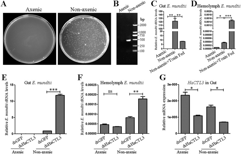
Fig 5. HaCTL3 depletion leads to increased load of E. mundtii in the gut and hemolymph.
(A, B) Confirmation of elimination of gut bacteria by culturing gut homogenates on LB agar plates (A), and by conducting PCR analysis on gut homogenates using universal primers (16S-F and 16S-R) of bacterial 16S rRNA gene (B). (C, D) Quantification of gut E. mundtii (C) and hemolymph E. mundtii (D) in axenic and nonaxenic larvae, as well as B. thuringiensis toxin-fed larvae. (E, F) Quantification of gut E. mundtii (E) and hemolymph E. mundtii (F) in axenic and nonaxenic larvae treated with dsHaCTL3 or dsGFP. Quantification was by qPCR analysis using primers of E. mundtii-specific 16S rRNA gene. (G) RT-qPCR analyses confirming the depletion of HaCTL3 transcripts in gut. The bar represents mean ± SEM from three biological replicates. *0.01 < p < 0.05, **0.001 < p < 0.01, ***p < 0.001 (Student’s t-test).

