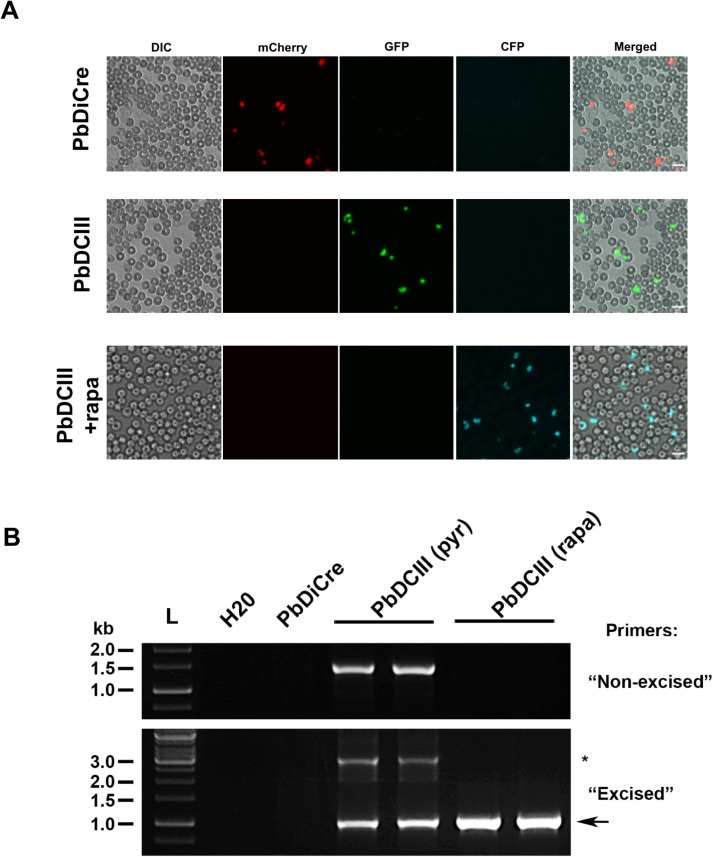
Fig 4. Exposure to rapamycin during the mammalian blood stages.
A. Fluorescence microscopy of unfixed blood stages in the parental PbDiCre parasites (upper panels) and in PbDCIII parasites before (middle panels) and after (lower panels) rapamycin treatment. Scale bar, 10 μm. B. PCR analysis of genomic DNA isolated from parental PbDiCre, pyrimethamine-selected (pyr) and rapamycin-treated (rapa) PbDCIII parasites, using primer combinations specific for non-excised or excised locus. A band corresponding to a non-excised locus can be amplified with the “excised” primer combination and is indicated with an asterisk.

