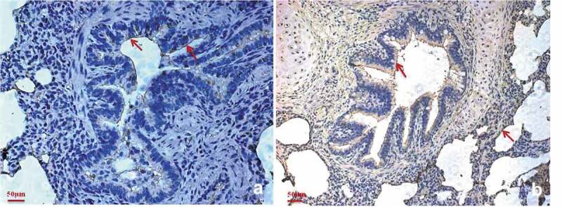
Figure 6.

Factor H is predominantly present in the M. hyopneumoniae colonization site.
Immunohistochemical staining of porcine bronchiole sections from M. hyopneumoniae-challenged pigs was performed using anti-factor H antibodies (a) or anti-P97 monoclonal antibodies (b). The presence of red staining shows the location of factor H (a) or M. hyopneumoniae strains (b). A widespread distribution of factor H can be observed in the bronchiole sections of all samples, especially in the ciliary borders of the bronchioles that are colocalized by M. hyopneumoniae.
