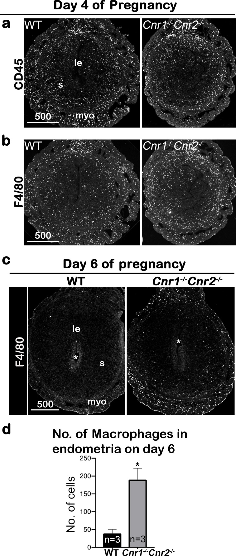
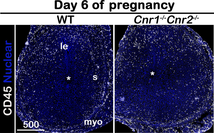
Figure 3. Macrophages are retained around implantation chambers of compromised deciduae in Cnr1-/-Cnr2-/- females.
(a) Uterine leukocytes in pre-implantation are highlighted by CD45, which is expressed on all leukocytes. (b) Immunofluorescent staining of F4/80 on day 4 of pregnancy to mark macrophages. (c) Immunofluorescent staining of F4/80 on day 6 of pregnancy. (d) The numbers of macrophages within the endometrial domains are quantified using three sections obtained from three different animals in each genotype. le, luminal epithelium; s, stroma; myo, myometrium; Asterisks, positions of embryos; Scale bars, 500 μm; *p<0.05, Student’s t-tests. All images are representative of three independent experiments.


