
Figure 5. Angiogenic factors are misregulated in Cnr1-/-Cnr2-/- and Cnr1-/- PDZ.
(a–d) In situ hybridization of Angpt1, Angpt2, Hif1a and Hif2a on day 6 of pregnancy. CK8 staining outlines uterine epithelial cells. (e and f) Immunostaining of HIF1A and HIF2A on day 6 of pregnancy. Dotted lines outline the PDZs. Asterisks, positions of embryos; Scale bars, 500 μm. All images are representative of three independent experiments.

Figure 5—figure supplement 1. Quantification of signals of angiogenic factors on day 6 of pregnancy.
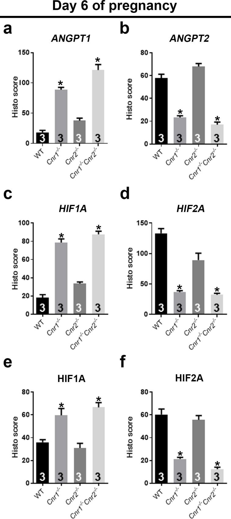
Quantification of in situ hybridization signals (a–d) and immunostaining signals (e and f) is plotted. H scores of signals in PDZs as outlined by dotted white lines are calculated for ANGPT1 in panel a and HIF1A in panels c and e. H scores for ANGPT2 signals are plotted in panel b. H scores for HIF2A signals are plotted in panels d and f. Values are mean ± SEM; Numbers on bars are sample sizes; *p<0.05, Unpaired t-tests with Welch’s correction.