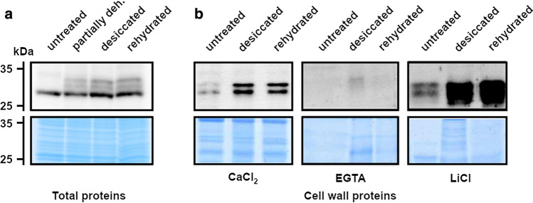
Fig. 3.
CpGLP1 protein expression. a Analysis of CpGLP1 protein accumulation was detected in proteins blots using a total proteins and b cell wall proteins. Total protein samples were prepared from untreated (100% relative water content; RWC), partially dehydrated (50% RWC), desiccated (2% RWC) and rehydrated (24 h) leaves. Cell wall proteins were prepared from 5 g using the same untreated, desiccated and rehydrated plant material used for total protein preparations. Briefly, plant material was subjected to sequential washes in increasing sucrose concentrations, and then cell wall proteins were extracted with CaCl2, EGTA and LiCl according to Printz et al. (2015). CaCl2, EGTA and LiCl fractions were concentrated by centrifugation and proteins were precipitated with acetone, resuspended in protein sample buffer and analyzed by 12% SDS-PAGE. Proteins on SDS gels were transferred to a nitrocellulose membrane for immunodetection or stained with Coomassie blue. Immunoblot analysis was performed with 1:5000 dilutions of the CpGLP1 antiserum

