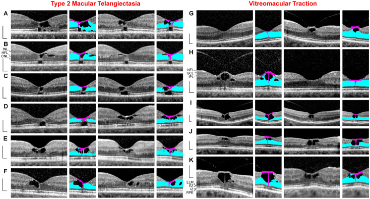
Fig. 1.
Müller cell cone provides the stability of the foveal center in cases of cystic disruption of the foveola. a–f Macular telangiectasia type 2. The images show linear scans through the fovea and parafovea in the left (left side) and right eyes (right side) of 6 patients. g–k Vitreomacular traction. The images show linear scans through the fovea and parafovea in the left (left side) and right eyes (right side) of 5 patients. The smaller images beside the scans show the Müller cell cone in pink and the Henle fiber (HFL)/outer nuclear layers (ONL) of the fovea in blue. Scale bars, 200 µm. ELM, external limiting membrane; EZ, ellipsoid zone; GCL, ganglion cell layer; INL, inner nuclear layer; IPL, inner plexiform layer; IZ, interdigitation zone; NFL, nerve fiber layer; OPL, outer plexiform layer; RPE, retinal pigment epithelium

