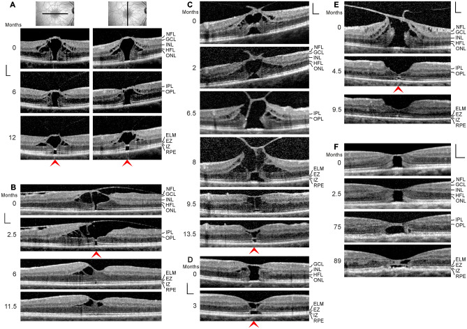
Fig. 4.
Regeneration of outer lamellar holes involves the formation of a hyperreflective glial tissue band which bridges the hole in the outer retina at the external limiting membrane (ELM) and the outer part of the outer nuclear layer (ONL) (arrowheads). The months after the first visit (0) are indicated left of the images. a A case of an outer lamellar hole. The orientations of the scans are shown above. b An outer lamellar hole which evolved into a tractional lamellar hole within 2.5 months after the first examination. Vitrectomy with internal limiting membrane (ILM) peeling was performed 2.6 months after the first visit. c Tractional generation and partial closure of an outer lamellar hole. Vitrectomy with ILM peeling was performed 8.1 months after the first visit. d Another outer lamellar hole. e Tractional generation and closure of an outer lamellar hole. Vitrectomy with ILM peeling was performed 0.5 month after the first visit. f Partial restoration of the contour of the central fovea after regeneration of the central ELM in an outer lamellar hole. Scale bars, 200 µm. EZ, ellipsoid zone; GCL, ganglion cell layer; HFL, Henle fiber layer; INL, inner nuclear layer; IPL, inner plexiform layer; IZ, interdigitation zone; NFL, nerve fiber layer; OPL, outer plexiform layer; RPE, retinal pigment epithelium

