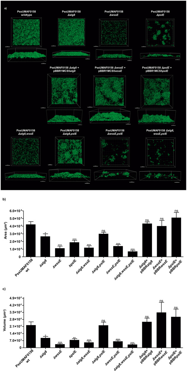
Fig. 4. Flow-cell chamber experiments of PssUMAF0158 wild-type and derived extracellular matrix mutants.
a Representative 48 h 3D biofilm images of GFP-tagged PssUMAF0158 wild-type and mutants are shown. The obtained images were analysed with the Leica Application Suite (Mannheim, Germany) and the IMARIS software package (Bitplane, Switzerland). Scale bar 20 µm. b Area in the field of view covered by 48 h biofilms of the GFP-tagged PssUMAF0158 wild-type, extracellular matrix mutants and complemented strains. c Volume in the field of view occupied by 48 h biofilms of the GFP-tagged PssUMAF0158 wild-type, extracellular matrix mutants and complemented strains. The area and volume values were calculated with the IMARIS software package (Bitplane, Switzerland). The following GFP-tagged strains were tested: PssUMAF0158 wild-type (PssUMAF0158 wt), PssUMAF0158 alginate mutant (Δalg8), PssUMAF0158 cellulose mutant (ΔwssE), PssUMAF0158 Psl-like polysaccharide mutant (ΔpslE), PssUMAF0158 Δalg8,wssE double mutant (Δalg8,wssE), PssUMAF0158 Δalg8,pslE double mutant (Δalg8,pslE), PssUMAF0158 ΔwssE,pslE double mutant (ΔwssE,pslE), PssUMAF0158 Δalg8,wssE,pslE triple mutant (Δalg8,wssE,pslE), alginate complemented strain (Δalg8 + pBBRalg8), cellulose complemented strain (ΔwssE + pBBRwssE) and Psl-like complemented strain (ΔpslE + pBBRpslE). A minimal of three replicates, and three independent experiments were performed. Statistical significance was assessed by two-tailed Mann–Whitney test (*p < 0.05, **p < 0.01, ***p < 0.001). Error bars show the standard error of the mean (s.e.m.).

