Fig. 4.
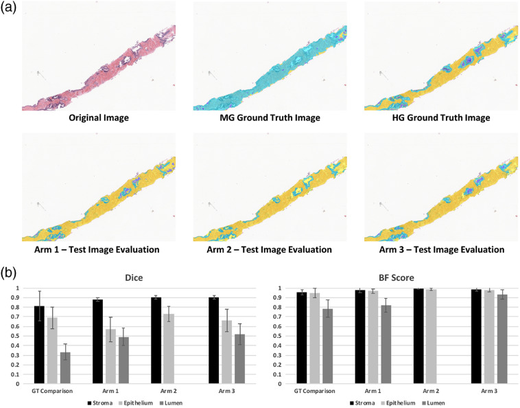
Evaluation of test set using individually trained SEL classification algorithms. (a) Using a biopsy sample that showed poor performance with the morphologically generated (MG) labeling, the fully trained segmentation algorithms (Arms 1 to 3) are compared to the MG ground truth and the HG ground truth. Background and foreground image regions were premasked for comparison. In all labeled images, stroma is labeled yellow, epithelium is labeled light blue, lumen is labeled dark blue. Note that the MG ground truth example shows noisy labeling compared to HG ground truth. Arm 1 shows a good balance between lumen and epithelium labeled, although many glands are left with incomplete epithelium label. Arm 2 shows good epithelium labeling; however, lumen label is almost completely missing. Arm 3 shows good balance between both models (Arm 1 and Arm 2). (B) Dice and BF Score comparison between the ground truth datasets (MG and HG).

