
Figure 3.
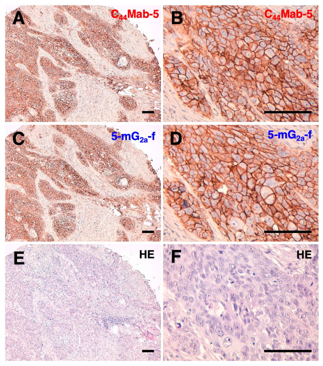
Immunohistochemical analysis using anti-CD44 mAbs against oral squamous cell carcinomas (OSCCs). (A and B) Consecutive tissue sections of OSCC were incubated with 1 µg/ml of C44Mab-5 for 1 h at room temperature followed by treatment with an Envision+ kit for 30 min. Color was developed using DAB for 2 min, and sections were then counterstained with hematoxylin. (C and D) Consecutive tissue sections of OSCC were incubated with 1 µg/ml of 5-mG2a-f for 1 h at room temperature followed by treatment with an Envision+ kit for 30 min. Color was developed using DAB for 2 min, and sections were then counterstained with hematoxylin. (E and F) Hematoxylin and eosin (HE) staining of consecutive tissue sections of OSCC. Scale bar, 100 µm. mAbs, monoclonal antibodies.