
Figure 8.
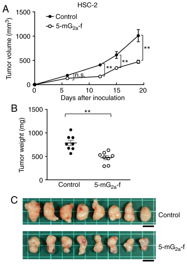
Evaluation of antitumor activity of 5-mG2a-f (from day 1) in HSC-2 ×enografts. (A) HSC-2 cells (5×10⁶ cells) were injected subcutaneously into the left flank. After day 1, 100 µg of 5-mG2a-f and control mouse IgG in 100 µl PBS were injected i.p. into treated and control mice, respectively. Additional antibodies were then injected on days 7 and 14. Tumor volume was measured on days 6, 12, 15, and 19. Values are mean ± SEM. Asterisk indicates statistical significance (**P<0.01; n.s., not significant, ANOVA and Sidak's multiple comparisons test). (B) Tumors of HSC-2 ×enografts were resected from 5-mG2a-f and control mouse IgG groups. Tumor weight on day 19 was measured from excised xenografts. Values are mean ± SEM. Asterisk indicates statistical significance (**P<0.01, Welch's t test). (C) Resected tumors of HSC-2 ×enografts from 5-mG2a-f and control mouse IgG groups on day 19. Scale bar, 1 cm.