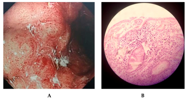
Figure 1.
Gastric lesion observed. The panel (A) shows endoscopic image obtained by conventional white light imaging in the patient’s stomach. An extended protruding lesion spanning over the greater curvature from the corpus to the antrum of stomach was noted, with irregular and reddened surface, which was bleeding easily on contact. The margin area of the lesion was saw-toothed onto a background mucosa marked with redness. The panel (B) shows a histological image obtained by microscopic examination (20×). This fragment of gastric mucosa was lined by dysplastic foveolar type epithelium, with a lamina propria exhibiting a poorly differentiated diffuse neoplasm and signet cells (>50%).

