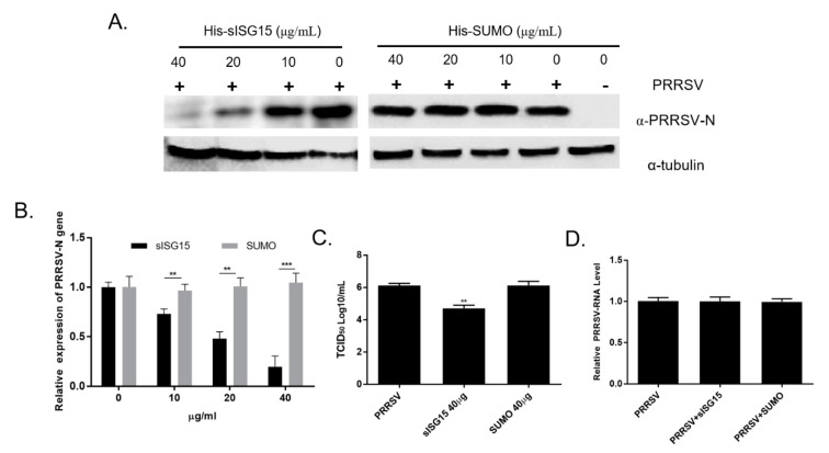
Figure 5.
Extracellular ISG15 inhibits PRRSV infection in PAMs. (A) PAMs were treated with various dilutions of recombinant sISG15 for 12 h then infected with the PRRSV-SD16 strain at 1 MOI. After 24 h, PAMs were harvested for Western blot analysis then blots were probed with homemade anti-PRRSV-N Mab-6D10 to monitor replication of PRRSV. PAMs treated with recombinant SUMO protein were included as a control. (B) PAMs were first treated with either sISG15 or SUMO protein for 12 h then were infected with PRRSV-SD16 strain at 1 MOI. After 24 h of infection, PAMs were harvested for qPCR evaluation of mRNA levels corresponding to PRRSV-N proteins. All experiments were repeated at least three times. Significant differences between indicated groups are marked by ** for p < 0.01 or *** for p < 0.01. (C) Cell culture supernatants from sISG15-treated or SUMO-treated PAMs followed by PRRSV-SD16 infection were titrated to quantify numbers of infectious virus particles in MARC-145 cells. All experiments were repeated at least three times. Significant differences between indicated groups are marked by ** for p < 0.01. (D) PAMs were pre-incubated with the 40 μg of either sISG15 protein or SUMO protein for 24 h at 37 °C or left untreated (control), followed by pre-chilling at 4 °C for 30 min prior to PRRSV inoculation. The PRRSV-JXA1 strain was used to inoculate the PAMs at 0.1 MOI at 4 °C for 1 h to avoid triggering of endocytosis. After washing cells using pre-chilled PBS to remove unbound virions, the attachment of PRRSV virions was evaluated using qPCR. All experiments were repeated at least three times.

