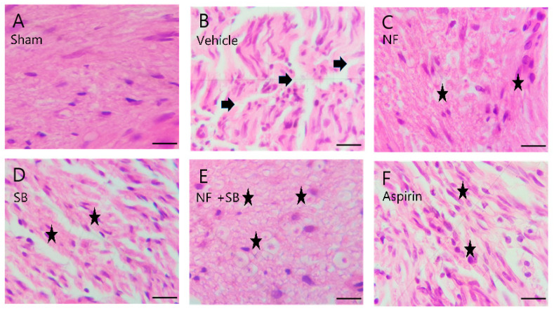
Figure 6.
Representative H&E staining of sciatic nerves on 14th day after surgery. (A) Sham group shows well-organized sciatic nerve fibers and no structure defects. (B) Vehicle group shows several areas of edema and degraded myelin sheath (arrows). (C) NF group, (D) SB group, (E) NF + SB group, (F) aspirin group, (C–F) The nerve fiber density was increased and the vacuolar-like degeneration was decreased at the 14th day (asterisks). Scale bars = 10 µm.

