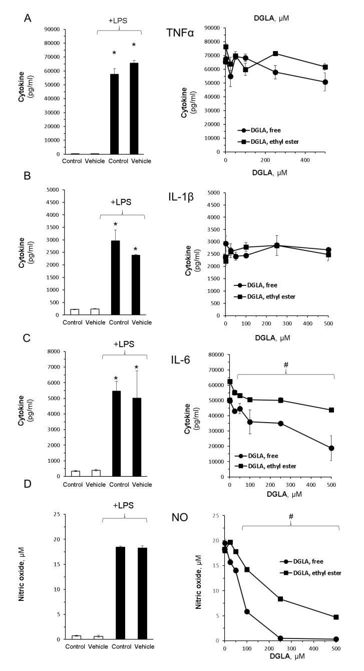
Figure 2.
DGLA modulates key inflammatory signals in bacterial lipopolysaccharide (LPS)-stimulated RAW264.7 macrophages. Inflammation was induced by LPS (100 ng/mL). Concomitantly, the cells were treated without or with increasing DGLA concentrations. DGLA was administered as free acid or ethyl ester form for 24 h. Tumor necrosis factor α (TNFα; (A)), interleukin 1β (IL-1β; (B)); interleukin 6 (IL-6; (C)), nitric oxide (NO; (D)), and were quantified in culture supernatants after 24 h. Data are presented as mean ± SD. */# denotes a significant difference compared to untreated control or LPS-stimulated control, respectively, p < 0.05, n = 3. Control—untreated cells; vehicle—cells treated with 0.1% DMSO, the solvent used in DGLA preparations. The left panel depicts the impact of LPS stimuli and lack of effect DMSO, whereas the right panel depicts the impact of increasing concentrations of DGLA on LPS-stimulated cells.

