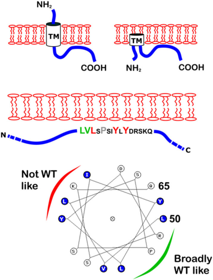
Figure 6.
Predicted topological models of E and the EPTM peptide. Above: Two of the suggested topologies of E are shown with TM, N- and C-termini indicated. Middle: The EPTM peptide is shown within the C-terminal region. Mutated residues are enlarged and coloured according to the level of membrane repartitioning observed. Below: A helical wheel depiction of the EPTM peptide with a suggested interpretation of sidedness with respect to membrane attachment.

