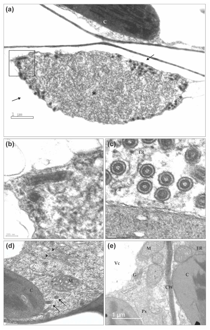
Figure 9.
Transmission electron micrographs of bean leaf infected by BaCV. (a) Overview of a viroplasm formed by coiled filamentous material (*) in the cytoplasm of a parenchymal cell. Typical rhabdovirus particles are present in the periphery of the viroplasm (arrows). (b) Details of the marked square with longitudinally-sectioned particles are depicted. (c) Cross-sections of BaCV particles show the internal and cylindrical nucleoprotein core, and the outer viral membrane, and also that virions are within a cavity of the endoplasmic reticulum. (d) Spongy parenchymal cell dually infected by BaCV and CPMMV. Brush-like aggregates of CPMMV particles (arrowheads) and BaCV in longitudinal and cross-sections (arrows) are visible. (e) Palisade parenchyma cells from a healthy bean plant. Chloroplast (C), endoplasmic reticulum (ER), Golgi complex (G), mitochondrion (M), peroxisome (Px), vacuole (Vc), and cell wall (CW).

