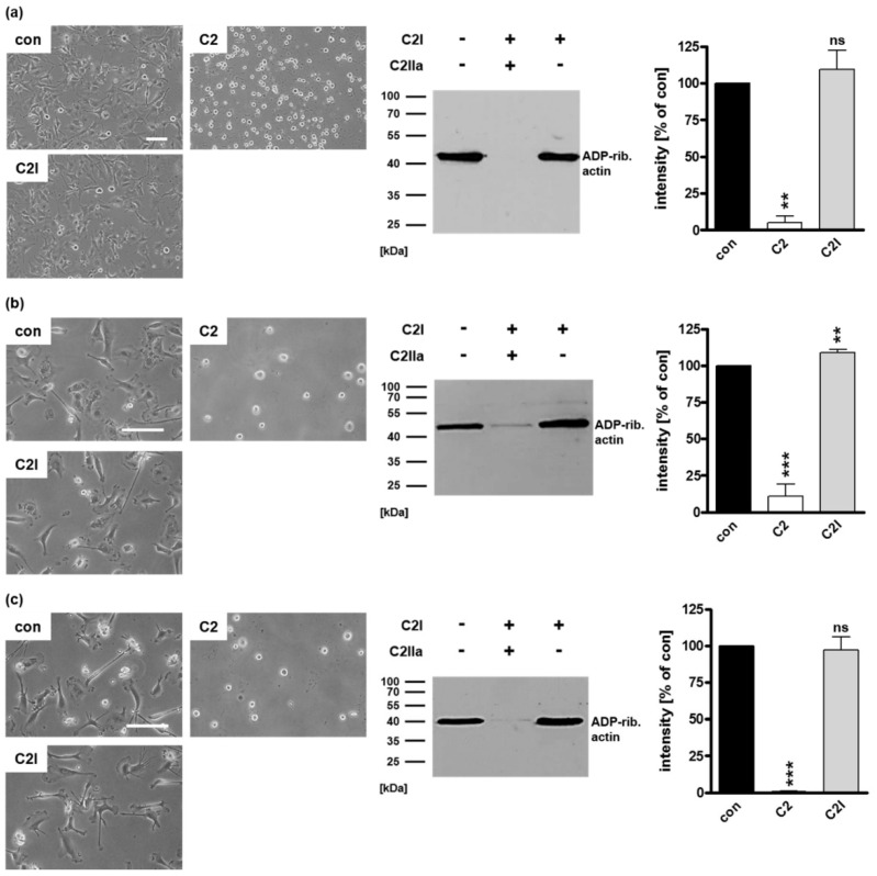
Figure 5.
The enzyme component C2I alone does not enter DCs. (a) U-DCS cells were incubated with C2I (80 nM), with complete C2 toxin (40/66 nM), or left untreated (con). After 5 h, representative phase contrast images were taken, cells were washed, lysed, and mixed with biotin-NAD+ (10 µM) and fresh C2I (300 ng) for 30 min at 37 °C. Subsequently, biotinylated, i.e., ADP-ribosylated actin (~40 kDa) was detected using peroxidase-coupled streptavidin in Western blot analysis. Equal protein loading was confirmed via Ponceau S staining of the membrane. Densitometrical analyses from two experiments (normalized to Ponceau S loading control) are given as mean ± SD. Immature (b) as well as mature (c) human monocyte-derived DCs were incubated with C2I (20 nM) without C2IIa, with complete C2 toxin (2/3.3 nM) or left untreated (con). After 5 h, representative phase contrast pictures are shown. Then, the cells were washed, lysed, and incubated with biotin-NAD+ (10 µM) and fresh C2I (300 ng) for 30 min at 37 °C. Next, Western blot detection of biotinylated, i.e., ADP-ribosylated actin (~40 kDa) was performed. Densitometrical analyses from three experiments (normalized to Coomassie-stained SDS-gel used as loading control) are given as mean ± SD. (a–c) Scale bars correspond to 50 µm and hold for all images. Significance was tested using a Student’s t test (ns = not significant, ** p < 0.01, *** p < 0.001).

